Abnormal Tendon on an MRI? Why It Might Not Mean Injury
This article may contain affiliate links. If you buy through them, we may earn a small commission at no extra cost to you. See our Affiliate Disclosure and Medical Disclaimer.
Here’s a scenario that trips up a lot of lifters and runners: you get an MRI or ultrasound for an unrelated issue, or out of curiosity, and the report comes back flagging “tendinosis,” “thickening,” or “structural abnormality” in a tendon that doesn’t actually hurt. It’s an unsettling thing to read. The honest, research-backed answer is that this is extremely common in people with zero symptoms, and a scan result by itself often isn’t the useful diagnostic tool it feels like it should be. Here’s what the actual imaging research says about the gap between what a tendon looks like and what it feels like.
At a Glance
| Question | What the Research Shows |
|---|---|
| How common are abnormal tendons in pain-free people? | Very — up to ~46% of asymptomatic runners show Achilles abnormalities in one study |
| Does an abnormal scan mean you have tendinopathy? | Not by itself — imaging and symptoms frequently don’t match |
| Does more activity mean a more abnormal-looking tendon? | Often yes — activity level is one of the strongest predictors of imaging findings |
| Should you get repeat scans to track a healing tendon? | Usually unnecessary once a diagnosis is established — it rarely changes treatment |
| Can a tendon look abnormal and never cause problems? | Yes, this is a well-documented and common pattern |
The Headline Number: Nearly Half of Runners Have “Abnormal” Achilles Tendons — With No Pain
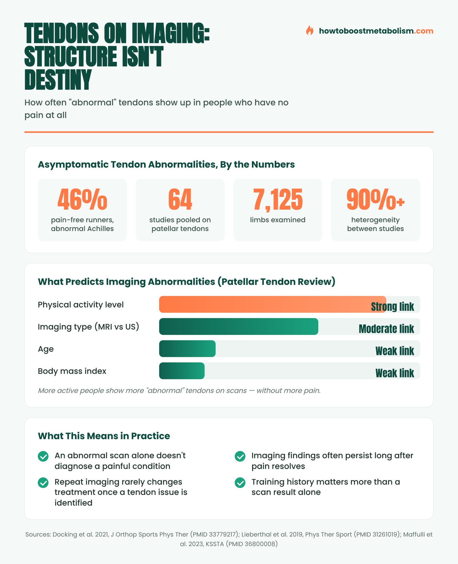
A cross-sectional study of 37 experienced male distance runners with no history of Achilles tendon pain, published in Physical Therapy in Sport (https://pubmed.ncbi.nlm.nih.gov/31261019/), used ultrasound imaging to check each runner’s tendons. Almost half — 46% — had at least one tendon flagged as abnormal on imaging, despite having never experienced Achilles pain. These weren’t borderline or ambiguous readings; they were classified as abnormal by the same criteria used to diagnose symptomatic tendinopathy.
This isn’t an isolated finding. A separate ultrasound study following 138 asymptomatic half- and full-marathon runners (276 tendons total) for 12 months after a race (https://pubmed.ncbi.nlm.nih.gov/37550955/) found specific abnormal patterns — focal areas of altered tissue echo — in a meaningful share of pain-free tendons before the race even started. Some of those patterns did predict who later developed pain, which is a genuinely useful finding, but plenty of runners with abnormal-looking tendons at the start of the study never developed symptoms at all during the follow-up year.
Free download
🔥 The Metabolism Starter Kit
3 honest guides: a 7-day reset plan, a foods cheat-sheet and 12 metabolism myths debunked. No spam — unsubscribe anytime.
It’s Not Just Runners, and Not Just the Achilles
A 2021 systematic review and meta-analysis in the Journal of Orthopaedic & Sports Physical Therapy pooled 64 studies covering 7,125 tendons from 4,616 people, specifically looking at patellar tendon abnormalities in people with no current or past tendon pain (https://pubmed.ncbi.nlm.nih.gov/33779217/). The studies varied enormously in what percentage of people showed abnormalities — so much so that the researchers couldn’t even calculate one overall summary number, because the heterogeneity between studies exceeded 90%.
What did reliably predict a higher chance of an “abnormal” finding, across that pooled data? Physical activity level and which imaging method was used (MRI detected more abnormalities than ultrasound) — not age, not body mass index, and not sample size. In plain terms: the more active and athletic someone is, the more likely their tendon is to look structurally different on a scan, completely independent of whether it hurts. An older-looking study of professional pallapugno (a traditional Italian throwing sport) players found a similar pattern — roughly a third to over half of asymptomatic throwers showed patellar tendon abnormalities on ultrasound (https://pubmed.ncbi.nlm.nih.gov/29387638/), and only a small fraction of those with abnormalities went on to develop actual symptoms over six months of follow-up.
Why Imaging and Symptoms Keep Disagreeing
A 2023 commentary in Knee Surgery, Sports Traumatology, Arthroscopy, co-authored by a leading tendon researcher, put the disconnect plainly (https://pubmed.ncbi.nlm.nih.gov/36800008/): recovery from pain and restoration of function “does not go hand in hand with the appearance of the affected tendon at imaging.” A tendon can remain structurally altered on a scan for a long time — possibly permanently — after someone’s pain has fully resolved and they’ve returned to full training. The authors go further, stating that the predictive value of abnormal findings in people without symptoms “remains limited,” and that treating an asymptomatic abnormal finding is “inappropriate and not supported by current evidence.”
This lines up with a broader pattern seen elsewhere in sports medicine and orthopedics — similar disconnects between structural imaging findings and actual symptoms show up with spinal disc abnormalities and knee cartilage changes in other research literatures. Tendons appear to follow that same rule: structure is one input into the pain experience, not a direct readout of it. Tendon tissue adapts to load over time, and some of that adaptation looks, on a scan, similar to early-stage pathology — without functioning like pathology at all.
Free download: our Metabolism Starter Kit — a protein calculator, a 7-day meal framework, and a one-page supplement checklist, with zero hype.
Get the Free Starter Kit →Why This Matters If You Get an Incidental Finding
The practical risk of not knowing this research is overreacting to a report you weren’t expecting. Getting imaging for an unrelated issue (a knee MRI that happens to note patellar tendon changes, for instance) and reading words like “tendinosis” or “structural abnormality” can trigger genuine anxiety, unnecessary activity restriction, or a cascade of follow-up scans — for a finding that may have been there, asymptomatically, for years and may never cause a problem.
The flip side matters too: if you do have genuine tendon pain that’s affecting your training, imaging findings alone shouldn’t be dismissed either. The point isn’t that tendon imaging is useless — it’s a genuinely useful tool when paired with your actual symptoms, training history, and a clinical exam. The point is that a scan result divorced from how the tendon actually feels and functions during real loading tells you much less than the report’s technical language implies.
A Practical Way to Interpret an Abnormal Finding
- Match the finding to your symptoms, not the other way around. If a tendon doesn’t hurt and functions normally under load, an incidental “abnormal” label on a scan taken for an unrelated reason usually doesn’t require treatment on its own.
- Repeat imaging rarely changes management once an initial diagnosis has been made — according to the 2023 KSSTA commentary, additional scans are “unlikely to provide further information or change prognosis” for an already-identified tendinopathy.
- Activity level predicts abnormal findings better than pain does. If you train hard, expect your tendons may look different on a scan than a sedentary person’s — that’s closer to an adaptation marker than a red flag by itself.
- A clinical exam and loading test matter more than the report’s wording. How a tendon responds to a hop, a single-leg heel raise, or a specific loading test tells a clinician more about function than the imaging description alone.
- If you do have pain, the finding is one piece of a bigger picture — training load, recent volume changes, and biomechanics usually explain more about why a tendon became symptomatic than the scan result does on its own.
A Closer Look at the Patellar Tendon Data
The 2017 ultrasound study of professional pallapugno throwers is worth sitting with for a moment because it’s small but unusually well followed-up (https://pubmed.ncbi.nlm.nih.gov/29387638/). Qualitative assessment found subclinical tendinopathy signs in 5 of 14 throwers’ non-dominant tendons (36%), while a separate, more sensitive quantitative measurement found structural abnormalities in 8 of 14 (57%). After six months of follow-up, only 2 of those 8 players with abnormal findings — 25% — went on to develop an actual clinical tendinopathy. The other 75% kept training and competing with a structurally “abnormal” tendon that never became a problem.
That 25% conversion rate is itself informative: having an abnormal-looking tendon on a scan does appear to raise your odds of eventually developing symptoms compared to a tendon that looks normal — it’s not a meaningless finding. But it’s also nowhere close to a guarantee, and three out of four athletes with the same scan result in this small study had no problem at all. That’s the nuance that gets lost when an abnormal finding gets reported to someone without that context: it’s a risk marker worth being aware of, not a diagnosis of an active injury.
How This Connects to Strength Training After 40
This matters more, not less, as training histories get longer. Someone who has lifted, run, or played sport consistently for 20+ years has had two decades of tendon loading and adaptation — exactly the kind of history the research above associates with a higher chance of an “abnormal-looking” tendon on a scan, independent of pain. If you’ve trained for most of your adult life and an incidental scan flags a tendon finding, your training history alone is a more parsimonious explanation than an assumption that something has suddenly gone wrong.
The practical takeaway for lifters over 40 fits the same pattern that runs through a lot of this site’s tendon coverage: structural findings and training history need to be read together, pain and function are the signals that actually drive decisions, and a label on a report is a starting point for a conversation with a clinician, not a verdict to act on alone.
What This Doesn’t Mean
None of this is a reason to ignore new or worsening tendon pain, swelling, or a sudden change in how a tendon feels under load — those are symptom-based signals, not imaging-based ones, and they still warrant attention. This research is specifically about the gap between a structural label on a report and the lived experience of pain and function. It’s a reason for proportionate interpretation of an unexpected finding, not a reason to dismiss a tendon problem you can actually feel.
If your scan looks worse than you feel, the practical question is how to keep training without flaring things up. A program built on moderate loads and controlled tempo — like Old School New Body‘s F4X method — is a more tendon-friendly way back to consistent lifting than chasing heavy singles. One honest caveat: ignore the age-related statistics on its sales page — they aren’t sourced — and judge the program by its structure, not the copy.
A joint- and tendon-friendly way to keep lifting:
Check Availability →Affiliate link — we may earn a commission at no extra cost to you. Disclosure
Frequently Asked Questions
Is it normal to have an abnormal-looking tendon with no pain?
Yes — research puts this at close to half of pain-free runners for the Achilles tendon, and similarly common rates have been found for the patellar tendon in other athletic populations. It’s a well-documented, common finding, not a rare anomaly.
Does an abnormal MRI or ultrasound mean I have tendinopathy?
Not by itself. Imaging findings and actual symptoms frequently don’t match. A tendon diagnosis should combine the imaging with your actual symptoms, training history, and a functional/loading exam.
Why do more active people have more tendon abnormalities on scans?
Tendon tissue adapts to repeated loading over time, and some of that adaptation can look structurally similar to early pathology on imaging without functioning like an injury. Physical activity level is one of the strongest predictors of imaging findings in research on asymptomatic athletes.
Should I get repeat scans to track how my tendon is healing?
Usually not necessary once an initial tendon diagnosis is established. Research suggests additional imaging is unlikely to provide new information or change treatment and prognosis in most cases.
What should I actually do if I get an unexpected abnormal tendon finding?
If the tendon doesn’t hurt and functions normally, discuss the finding with a clinician in the context of your symptoms and activity level rather than treating the report’s wording as a diagnosis on its own. If it does hurt, get it properly assessed rather than self-diagnosing from the report.
For related reading on managing tendons that do hurt, see our guides to eccentric training for tendon health, isometric training for joint pain, and tennis elbow in lifters. If grip and finger tendons are your concern specifically, our deep dive on finger pulley injuries covers a related “injury or adaptation” imaging finding in climbers.
This article is for educational purposes and is not medical advice. Imaging reports should always be interpreted by a qualified clinician in the context of your specific symptoms and history — do not self-diagnose or change your activity based on a report alone. See our full Medical Disclaimer.